Publications
Selected Publications
⭐Key publications
Enhancing Retinal Stem Cells Survival In Vivo
Bhargava M, Mehta KN, Parikh BH, Liu Z, Su X.
Stem Cell Res Ther. 2026 Mar 22. doi: 10.1186/s13287-026-04971-0.
Soundararajan L, Surendran H, Patlolla N, Battu R, Stoddard J, Arrizabalaga S, Liu Z, Lingam G, Su X, Ryals RC, Pal R.
NPJ Regen Med. 2025 Apr 19;10(1):19. doi: 10.1038/s41536-025-00407-0.
Liu H, Huang SS, Lingam G, Kai D, Su X, Liu Z.
Stem Cell Res Ther. 2024 Oct 31;15:390. doi: 10.1186/s13287-024-04007-5.
Single-cell transcriptomics reveals maturation of transplanted stem cell-derived retinal pigment epithelial cells toward native state
Parikh BH, Blakeley P, Regha K, Liu Z, Yang B, Bhargava M, Wong DSL, Tan QSW, Wong CSW, Wang HF, Al-Mubaarak A, Chou C, Cheung CMG, Lim KL, Barathi VA, Hunziker W, Lingam G, Hu TX, Su X
Proc Natl Acad Sci U S A. 2023 Jun 27;120(26):e2214842120.
doi: 10.1073/pnas.2214842120.
Stem cell-derived retinal pigment epithelial (RPE) cell transplantation represents a promising therapeutic strategy for age-related macular degeneration, yet the transcriptional changes that occur in transplanted cells within the host retinal environment have remained poorly characterised. Published in PNAS, this study employed single-cell RNA sequencing to profile stem cell-derived RPE monolayers retrieved from immunocompetent rabbit eyes 30 days after subretinal transplantation, benchmarked against age-matched in vitro counterparts. Transplanted RPE cells — derived independently from both embryonic and induced pluripotent stem cell sources — demonstrated a consistent, unidirectional maturation trajectory toward the native adult human RPE transcriptional state, accompanied by upregulation of genes associated with extracellular matrix organisation, oxidoreductase activity, and lipid metabolism. Gene regulatory network analysis identified three transcription factors, FOS, JUND, and MAFF, specifically activated in post-transplanted RPE, with downstream targets implicated in photoreceptor support and cell survival. These findings provide a molecular framework for evaluating RPE transplant outcomes at single-cell resolution, with implications for the optimisation and clinical translation of cell-based therapies for retinal degeneration.
Regha K, Bhargava M, Al-Mubaarak A, Chai C, Parikh BH, Liu Z, Wong CSW, Hunziker W, Lim KL, Su X.
Sci Rep. 2022 Sep 12. doi: 10.1038/s41598-022-19777-2.

cGMP-grade human iPSC-derived retinal photoreceptor precursor cells rescue cone photoreceptor damage in non-human primates
Lingam S, Liu Z, Yang B, Wong W, Parikh BH, Ong JY, Goh D, Wong DSL, Tan QSW, Tan GSW, Holder GE, Regha K, Barathi VA, Hunziker W, Lingam G, Zeng X, Su X*.
Stem Cell Res Therapy. 2021 Aug 19;12(1):463. doi: 10.1186/s13287-021-02539-8.
Addressing the critical need for regenerative treatments for end-stage inherited retinal diseases, our study in Stem Cell Research & Therapy evaluates the therapeutic efficacy of clinical-grade (cGMP) human induced pluripotent stem cell (iPSC)-derived photoreceptor precursors. By utilizing a non-human primate model, we demonstrated that sub-retinally transplanted cell suspensions are highly biocompatible and capable of surviving and maturing into functional cone photoreceptors over a three-month period. A key highlight of this research is the observed structural recovery of the retinal ellipsoid zone in damaged models, providing measurable evidence of the cells’ ability to rescue retinal integrity. Beyond the biological success, this work proves the feasibility of using US FDA-approved, cryopreserved cell lines for international clinical applications. These findings offer a validated, scalable roadmap for transitioning cell replacement therapies into the clinic, aligning with the highest safety and manufacturing protocols.
Liu Z, Ilmarinen T, Tan GSW, Hongisto H, Wong EYM, Tsai ASH, Al-Nawaiseh S, Holder GE, Su X, Barathi VA, Skottman H, Stanzel BV.
Stem Cell Res Ther. 2021 Jul 27;12(1):423. doi: 10.1186/s13287-021-02395-6.
Seah I, Liu Z, Soo Lin Wong D, Wong W, Holder GE, Amutha Barathi V, Lingam G, Su X, Stanzel BV.
J Vis Exp. 2021 Jun 14;(172). doi: 10.3791/62638.
Liu Z, Parikh BH, Tan QSW, Wong DSL, Ong KH, Yu W, Seah I, Holder GE, Hunziker W, Tan GSW, Barathi VA, Lingam G, Stanzel BV, Blenkinsop TA, Su X.
Stem Cell Reports. 2021 Feb 9;16(2):237-251. doi: 10.1016/j.stemcr.2020.12.007.
Discovering Retinal Therapeutics
Modeling inherited retinal diseases using human induced pluripotent stem cell derived photoreceptor cells and retinal pigment epithelial cells
Seah I, Goh D, Banerjee A, Su X
Front Med (Lausanne). 2024 Jul 1;11:1328474. doi: 10.3389/fmed.2024.1328474.
Inherited retinal diseases (IRDs) represent a significant group of untreatable disorders where progress is often hindered by the lack of biologically relevant human tissue models, In this comprehensive review published in Frontiers in Medicine, we provide a detailed toolkit for leveraging human induced pluripotent stem cell (iPSC) and CRISPR/Cas9 technologies to create patient-specific 2D cellular cultures and 3D retinal organoids. We explore critical considerations for establishing robust models—including somatic cell sources, isogenic control strategies, and quality control metrics—while dissecting current iPSC-based models for conditions such as Retinitis Pigmentosa and Leber congenital amaurosis. This work underscores how high-fidelity, human-centric platforms can revolutionize drug discovery and precision medicine. By aligning with the FDA Modernization Act 2.0, these advanced in vitro models offer a scalable and scientifically superior alternative to traditional animal testing, paving the way for more effective target validation and the development of transformative ocular therapies
Liu H, Wu C, Hu S, Leng B, Lou X, Liu Z, Su X, Huang D
J Agric Food Chem. 2024 Jul 3;72(26):14701-14712. doi: 10.1021/acs.jafc.4c01537.
Hu W, Zheng R, Feng Y, Tan D, Chan Chung-Tsing G, Su X, Kim JE
Food Funct. 2023 Sep 19;14(18):8321-8330. doi: 10.1039/d3fo02105j.
Wong WM, Sun W, Vyas C, Fong AHC, Chee CK, Su X, Teo KY, Cheung CMG
Br J Ophthalmol. 2023 May;107(5):698-704. doi: 10.1136/bjo-2022-322457.
Lenin RR, Koh YH, Zhang Z, Yeo YZ, Parikh BH, Seah I, Wong W, Su X
Int J Mol Sci. 2023 May 15;24(10):8763. doi: 10.3390/ijms24108763.
Lim SY, Wong WM, Seah I, Chan HW, Su X, Lingam G, Yuen YS
Graefes Arch Clin Exp Ophthalmol. 2023 Feb;261(2):303-315. doi: 10.1007/s00417-022-05770-y.
Wu KX, Yeo NJY, Ng CY, Chioh FWJ, Fan Q, Tian X, Yang B, Narayanan G, Tay HM, Hou HW, Dunn NR, Su X, Cheung CMG, Cheung C
BMC Biol. 2022 Feb 15;20(1):47. doi: 10.1186/s12915-022-01244-z.
Seah I, Goh D, Chan HW, Su X
Genes. 2022 Feb 14;13(2):344. doi: 10.3390/genes13020344
Tan TE, Fenner BJ, Barathi VA, Tun SBB, Wey YS, Tsai ASH, Su X, Lee SY, Cheung CMG, Wong TY, Mehta JS, Teo KYC
Frontiers in Genetics. 2021 Dec 7;12:795010. doi: 10.3389/fgene.2021.795010
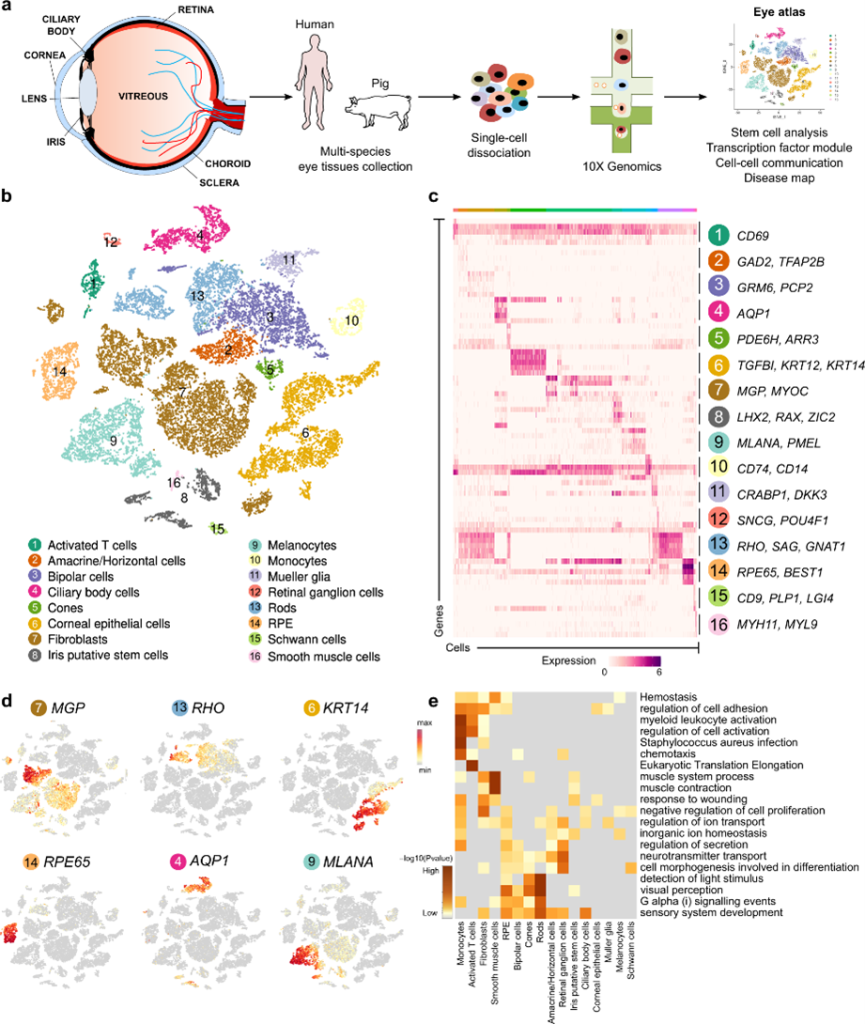
Multi-species single-cell transcriptomic analysis of ocular compartment regulons
Gautam P, Hamashima K, Chen Y, Zeng Y, Makovoz B, Parikh BH, Lee HY, Lau KA, Su X, Wong RCB, Chan WK, Li H, Blenkinsop TA, Loh YH.
Nature Communication. 2021 Sep 28;12(1):5675. doi: 10.1038/s41467-021-25968-8
Our research, published in Nature Communications, presents the first comprehensive multi-species single-cell transcriptomic atlas of the eye, mapping the cornea, iris, ciliary body, neural retina, RPE, and choroid. By integrating data across humans, pigs, mice, and zebrafish, we identified conserved and species-specific transcriptional regulons that govern cellular identity and function. A significant highlight of this work is the discovery of putative adult stem cell populations within the iris tissue, which exhibits high stem-cell potency and potential for regenerative applications. Furthermore, we established a high-resolution disease map and a viral-entry map—identifying receptors for pathogens like SARS-CoV-2—to provide a foundational resource for understanding ocular disorders across different compartments. This atlas offers a powerful roadmap for identifying novel therapeutic targets and understanding the molecular drivers of cell maturation, as demonstrated by our findings on the role of the transcription factor KLF7 in retinal ganglion cell development.
Wong WM, Chee C, Bhargava M, Chai C, Lin H, Zhao P, Ariadarma Mangunkusumo E, Naing T, Yuen YS, Wong TY, Su X, Lingam G
J Ophthalmol. 2020 Mar 19;2020:1875860. doi: 10.1155/2020/1875860.
Su X, Tan QS, Parikh BH, Tan A, Mehta MN, Sia Wey Y, Tun SB, Li LJ, Han XY, Wong TY, Hunziker W, Luu CD, Owada Y, Barathi VA, Zhang SS, Chaurasia SS
Invest Ophthalmol Vis Sci. 2016 Jun 1;57(7):3397-408. doi: 10.1167/iovs.15-18542
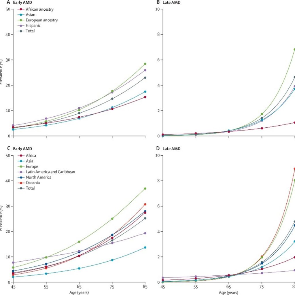
Global prevalence of age-related macular degeneration and disease burden projection for 2020 and 2040: a systematic review and meta-analysis
Wong WL*, Su X*, Li X, Cheung CMG, Klein R, Cheng CY, Wong TY
Lancet Glob Health. 2014 Feb;2(2):e106-16. doi: 10.1016/S2214-109X(13)70145-1.
Age-related macular degeneration (AMD) is a leading cause of blindness worldwide, yet comprehensive global estimates of its burden have been lacking. In this systematic review and meta-analysis published in The Lancet Global Health, we analysed data from 39 population-based studies encompassing over 129,000 individuals to estimate AMD prevalence across ethnicities and geographic regions. Our findings show that approximately 8.7% of the global population is affected, with prevalence significantly higher in people of European ancestry compared to Asian and African populations. Using UN population projection data, we forecast that the number of people living with AMD will rise from 196 million in 2020 to 288 million by 2040, with Asia bearing the greatest absolute burden due to its large and rapidly ageing population. These estimates highlight a critical and growing global health challenge, particularly in regions where access to anti-angiogenic treatments remains limited. Our data provide an evidence base to inform eye care policy, resource allocation, and the design of targeted interventions at both regional and global levels.
Clinic
Wong WM, Tham YC, Simunovic MP, et al.
Asia Pac J Ophthalmol (Phila). 2024 Jan-Feb;13(1):100030. doi: 10.1016/j.apjo.2023.100030.
Lingam G, Su X
Indian J Ophthalmol. 2020 Jun;68(6):1100-1101. doi: 10.4103/ijo.IJO_16_20
Su X, Yin WT
Nature Communication
JAMA Ophthalmology. 2021 Dec 1;139(12):1307-1308. doi:10.1001/jamaophthalmol.2021.4602
Developing Tools for
Retinal Stem Cells Transplant
Retinal Stem Cells Transplant
Loh WW, Lin Q, Zhao X, Su X, Loh XJ, Lim JYC
Chem Asian J. 2024 Jun 15:e202400453. doi: 10.1002/asia.202400453.
Arwani RT, Tan SCL, Sundarapandi A, Goh WP, Liu Y, Leong FY, Yang W, Zheng XT, Yu Y, Jiang C, Ang YC, Kong L, Teo SL, Chen P, Su X, Li H, Liu Z, Chen X, Yang L, Liu Y
Nat Mater. 2024 Jun 12. doi: 10.1038/s41563-024-01918-9.
Zhang K, Liu Z, Lin Q, Boo YJ, Ow V, Zhao X, Wong DSL, Lim JYC, Xue K, Su X, Wu D, Loh XJ
Biomater Res. 2022 Dec 2. 26(1):70. doi:10.1186/s40824-022-00316-z
A bio-functional polymer that prevents retinal scarring through modulation of NRF2 signalling pathway
Parikh BH, Liu Z, Blakeley P, Lin Q, Singh M, Ong JY, Ho KH, Lai JW, Bogireddi H, Tran KC, Lim JYC, Xue K, Al-Mubaarak A, Yang B, R S, Regha K, Wong DSL, Tan QSW, Zhang Z, Jeyasekharan AD, Barathi VA, Yu W, Cheong KH, Blenkinsop TA, Hunziker W, Lingam G, Loh XJ, Su X
Nat Commun. 2022 May 19 ; 13:30474. doi: 10.1038/s41467-022-30474-6
Our research, published in Nature Communications, introduces poly(CEP), a synthetic bio-functional polymer designed to prevent retinal scarring, or proliferative vitreoretinopathy (PVR), a primary cause of failed retinal detachment surgeries. Challenging the traditional view of polymers as inert drug carriers, poly(CEP) acts as an active therapeutic agent by shedding micelles that are internalized by retinal cells via clathrin-dependent endocytosis to activate the NRF2 signaling pathway. This molecular mechanism effectively suppresses epithelial-mesenchymal transition (EMT), hyper-proliferation, and migration—the biological drivers of fibrosis. In experimental models, poly(CEP) demonstrated superiority over standard clinical treatments, such as SF6 gas, by maintaining retinal attachment and preventing contractile membrane growth without the need for adjunct pharmacological agents. These findings highlight a transformative approach in nanomedicine, where bio-engineered materials are specifically designed to modulate intracellular signaling to treat complex wound-healing disorders and other fibrotic diseases
Tan RPT, Cheng JJW, Parikh BH, Wong JHM, Soh BW, Chang JJ, Tran KC, Lee Y, Chee PL, Boo YJ, Lin Q, Jiang L, Su X, Lim JYC, Loh XJ, Xue K
ACS Appl Polym Mater. 2022 Jul;4(7):5091–5102. doi: 10.1021/acsapm.2c00614.
Anti-Angiogenic Nanomicelles for the Topical Delivery of Aflibercept to Treat Retinal Neovascular Disease
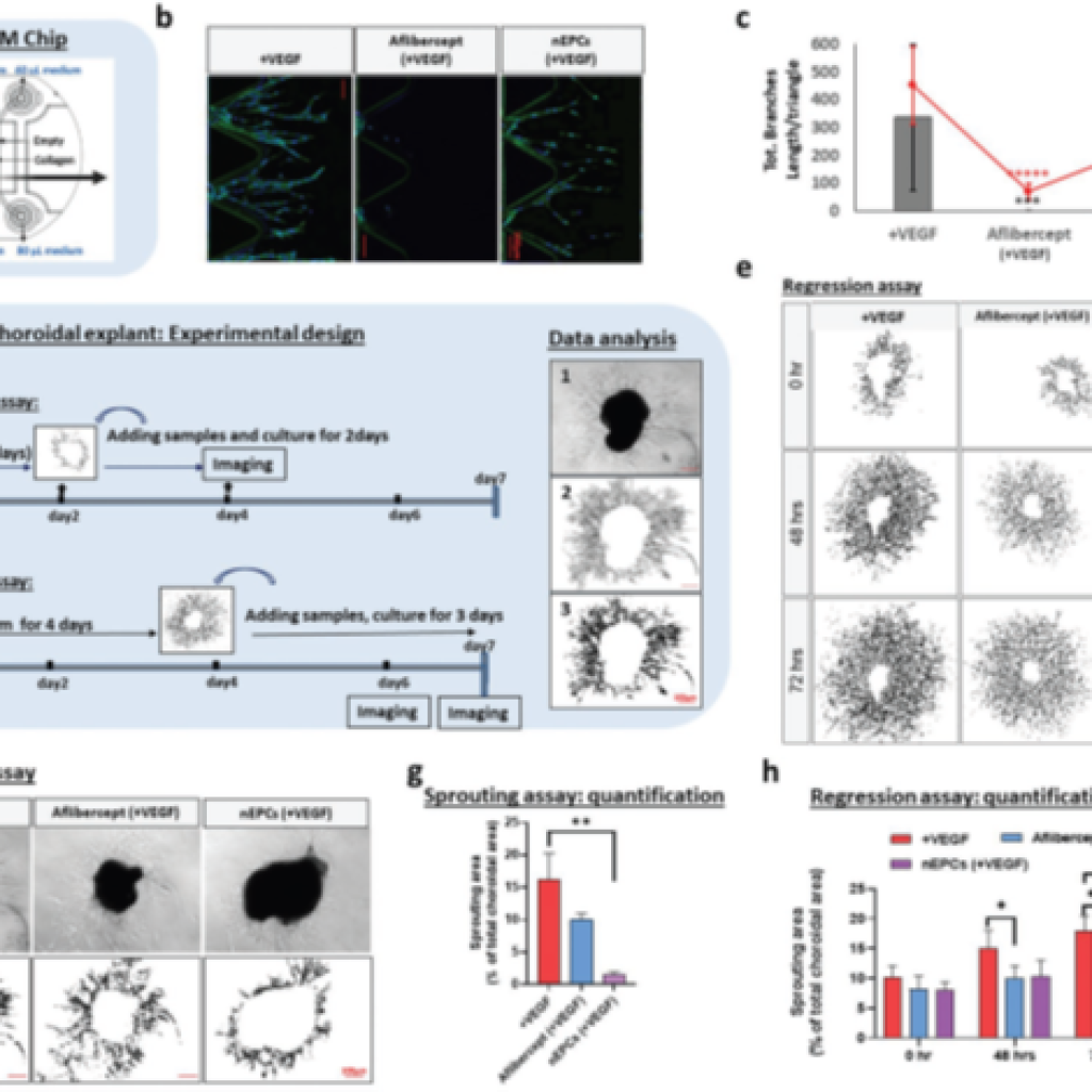
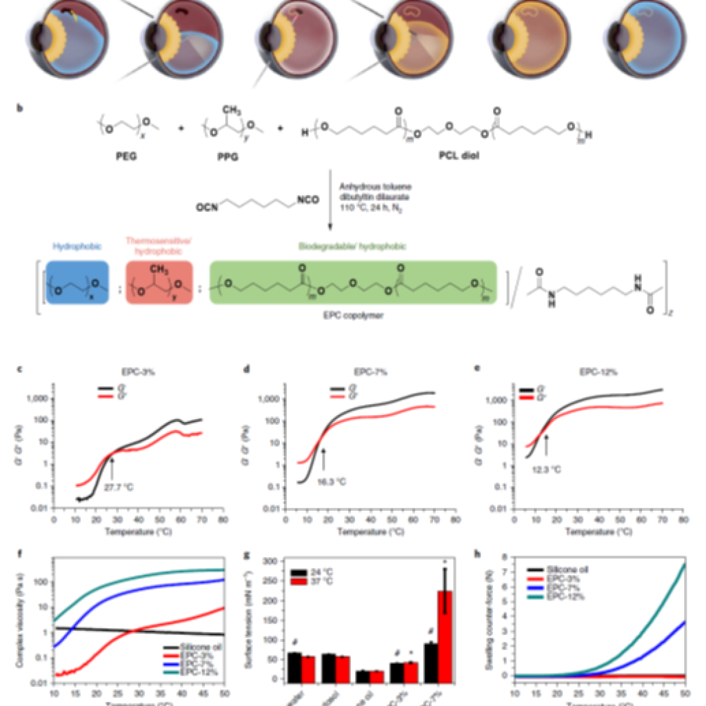
Zhao X, Seah I, Xue K, Wong W, Tan QSW, Ma X, Lin Q, Lim JYC, Liu Z, Parikh BH, Mehta KN, Lai JW, Yang B, Tran KC, Barathi VA, Cheong KH, Hunziker W, Su X, Loh XJ
Advanced Materials. 2021 Nov 2;e2108360. doi: 10.1002/adma.202108360
Intravitreal injection of anti-vascular endothelial growth factor (anti-VEGF) agents remains the standard of care for neovascular retinal diseases, yet the associated treatment burden and risk of sight-threatening complications underscore the need for less invasive delivery strategies. Published in Advanced Materials, this study reports the development of a polymeric nanomicelle system — composed of the triblock copolymer EPC (poly(ethylene glycol), poly(propylene glycol), and polycaprolactone) — for the topical delivery of aflibercept to the posterior segment of the eye. The nanomicelles demonstrated a 47.3% encapsulation efficiency for aflibercept, enhanced corneal penetration in ex vivo porcine models, and achieved vitreous drug concentrations above the clinically relevant IC₅₀ threshold following topical administration in murine models. In laser-induced choroidal neovascularisation models, topically applied nEPCs loaded with aflibercept produced measurable lesion regression. Notably, unloaded nEPCs also exhibited intrinsic antiangiogenic activity in vitro and ex vivo, modulating both VEGF and PDGF signalling pathways, suggesting potential synergistic effects with the encapsulated therapeutic.
Lin Q, Liu Z, Wong DSL, Lim CC, Liu CK, Guo L, Zhao X, Boo YJ, Wong JHM, Tan RPT, Xue K, Lim JYC, Su X, Loh XJ
Biomaterials. 2021 Nov 17;121262. doi: 10.1016/j.biomaterials.2021.121262
Seah I, Zhao X, Lin Q, Liu Z, Su SZZ, Yuen YS, Hunziker W, Lingam G, Loh XJ, Su X
Eye (Lond). 2020 Aug;34(8):1341–1356. doi: 10.1038/s41433-020-0770-y. Erratum in: Eye (Lond). 2020 May 22.
Xue K, Liu Z, Jiang L, Kai D, Li Z, Su X, Loh XJ
Biomater Sci. 2020 Feb 4;8(3):926-936. doi: 10.1039/c9bm01603a
Xue K, Zhao X, Zhang Z, Qiu B, Tan QSW, Ong KH, Liu Z, Parikh BH, Barathi VA, Yu W, Wang X, Lingam G, Hunziker W, Su X, Loh XJ
Biomater Sci. 2019 Nov 1;7(11):4603-4614. doi: 10.1039/c9bm01049a.
Retinal-detachment repair and vitreous-like-body reformation via a thermogelling polymer endotamponade
Liu Z, Liow SS, Lai SL, Alli-Shaik A, Holder GE, Parikh BH, Krishnakumar S, Li Z, Tan MJ, Gunaratne J, Barathi VA, Hunziker W, Lakshminarayanan R, Tan CWT, Chee CK, Zhao P, Lingam G, Loh XJ, Su X
Nat Biomed Eng. 2019 Aug;3(8):598-610. doi: 10.1038/s41551-019-0382-7.
Current internal tamponade agents used in vitreoretinal surgery function through buoyancy forces and are associated with complications including post-operative positional requirements, elevated intraocular pressure, cataract formation, and the need for surgical removal. Published in Nature Biomedical Engineering, this study describes a thermogelling polymer endotamponade that operates instead through surface tension and swelling counter-forces, offering a mechanistically distinct approach to retinal tamponade. Biocompatibility was assessed in rabbit vitrectomy models, with surgical efficacy further evaluated in a non-human primate model of retinal detachment. Notably, as the thermogel biodegrades over approximately three months post-surgery, it promotes the reformation of a vitreous-like body that recapitulates the biophysical properties of the native vitreous humour, suggesting utility not only as a surgical adjunct but as a candidate long-term vitreous substitute.
Chan SY, Chan BQY, Liu Z, Parikh BH, Zhang K, Lin Q, Su X, Kai D, Choo WS, Young DJ, Loh XJ
ACS Omega. 2017 Dec 31;2(12):8959-8968. doi: 10.1021/acsomega.7b01604.
Su X, Tan MJ, Li Z, Wong M, Rajamani L, Lingam G, Loh XJ
Biomacromolecules. 2015 Oct 12;16(10):3093-102. doi: 10.1021/acs.biomac.5b01091.